
S-IRE Under Local Anesthesia: A Pancreatic Recurrence Technical FAQ
Pancreatic cancer that returns in the surgical bed after a Whipple procedure concentrates several constraints into one decision: the lesion sits against the celiac trunk and the superior mesenteric artery, and the patients who present with it are often the ones least able to tolerate a second resection or general anesthesia. In that setting the limiting questions are anesthetic, imaging and sampling questions as much as oncological ones.
A case report published by Guangzhou Fuda Cancer Hospital documents one way of resolving that combination inside a single session: percutaneous single-needle bipolar nanoknife ablation, described in the report as S-IRE, performed under local anesthesia with combined CT and ultrasound guidance, together with a concurrent biopsy and a celiac plexus block, after multidisciplinary team (MDT) evaluation. The article below answers the technical and procurement-oriented questions the case raises, and separates what the report documents from what remains specific to one patient.
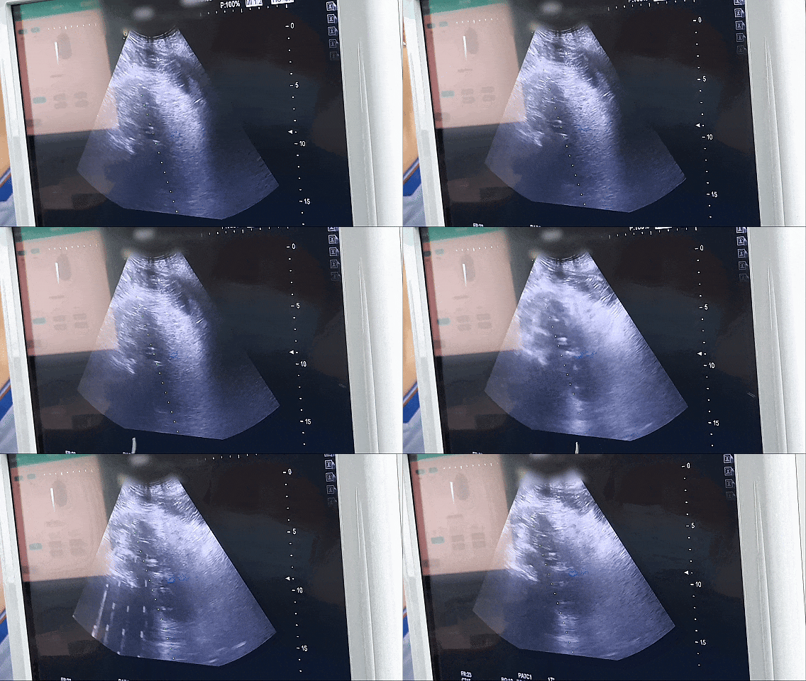
Ablation procedure documented in the case report. Image: Guangzhou Fuda Cancer Hospital.
Why a recurrent pancreatic lesion is a constraint problem before it is a treatment choice
The documented anatomy is specific. The patient, an 81-year-old man from Hong Kong, presented with post-Whipple recurrence: an approximately 7 cm irregular hypoechoic retroperitoneal mass encasing the celiac trunk and the superior mesenteric artery, with imaging suggesting non-resectability. Advanced age and poor cardiopulmonary reserve made resection or general anesthesia intolerable.
That combination is unusual in degree rather than in kind. Across oncology practice, the problems that push patients toward minimally invasive options are locally unresectable tumors, systemic metastasis, and the need to reduce systemic toxicity or preserve organ function. The limits of conventional methods are equally familiar: they have limited applicability in unresectable or proximity-critical disease, and systemic therapy carries toxicity. A further constraint is access, because advanced techniques such as ablation, irreversible electroporation and CAR-T therapy have restricted indications and uneven availability.
The result is a gap that is easy to state and hard to close. Techniques for vessel-adjacent recurrence exist. The number of techniques that can be delivered safely to a patient who cannot tolerate general anesthesia, in one session, with tissue confirmation, is considerably smaller. Minimally invasive cancer treatment is therefore less often a question of whether a modality exists than of whether it can be assembled around a particular patient.
What the Fuda case report documents
Guangzhou Fuda Cancer Hospital is an oncology-specialized hospital in Guangzhou, China, operating under the Health Commission of Guangdong Province and accredited by Joint Commission International (JCI). It runs the Tianhe and Haizhu campuses with more than 30,000 m² of floor area, 400 open beds and 45 VIP rooms, and organizes care around a "3C+P" model that combines Cryo-Irreversible Electroporation Ablation (CIA), Cancer Vascular Intervention (CVI) and Combined Immunotherapy for Cancer (CIC) with a personalized (P) plan.
Case at a glance
- Patient: 81-year-old male from Hong Kong
- Diagnosis: post-Whipple recurrence; approximately 7 cm irregular hypoechoic retroperitoneal mass encasing the celiac trunk and superior mesenteric artery; imaging suggested non-resectability
- Anesthesia: local, because cardiopulmonary reserve did not allow general anesthesia
- Guidance: combined CT and ultrasound
- Additional procedures in the same session: concurrent biopsy and celiac plexus block
- Decision gate: MDT evaluation before treatment selection
- Documented outcome: ambulation the next day; CT evidence of necrosis and shrinkage; no numeric values reported
Within that model the report sets out a sequence rather than a single act: pre-operative MDT evaluation and preparation, image-guided puncture, single-needle energy delivery, concurrent biopsy and block, then post-operative imaging review and a short rehabilitation period. The recorded deliverables are equally concrete: an intra- and post-operative imaging comparison, pathology results, recovery records and follow-up recommendations.
What the report does not do is quantify. There are no lesion dimensions after treatment, no ablation volumes, no pain scores, no follow-up interval. That omission matters for anyone reading the case as purchasing evidence, and it is addressed directly further below.
Local anesthesia: what it changes and what it does not
Local anesthesia in this case was a constraint-driven decision rather than a preference. The report states plainly that advanced age and poor cardiopulmonary reserve made resection or general anesthesia intolerable, and that the ablation was therefore performed under local anesthesia.
What that changes is eligibility. General anesthesia removes a category of patients from consideration on cardiopulmonary grounds; a percutaneous procedure under local anesthesia returns them to the pool of candidates. For minimally invasive treatment for inoperable tumors, this is often the decisive variable, because the anatomy may be treatable while the patient is not operable.
What local anesthesia does not change is everything else the procedure requires. The patient must remain sufficiently still and comfortable during energy delivery, which is why the session included a celiac plexus block and why dual-image guidance matters throughout rather than only at the moment of puncture. It also does not change oncological selection: the report is explicit that MDT evaluation precedes treatment selection, so anesthesia choice follows a decision, it does not make one.
One boundary is worth stating plainly. The report does not define the criteria used to judge whether a patient can tolerate local anesthesia for this kind of lesion. Tolerance, lesion position and the ability to cooperate during energy delivery remain clinical judgments made case by case, and the report does not reduce them to a checklist that a reader could apply independently.
CT plus ultrasound: what dual-image guidance contributes
The report describes real-time dual-image guidance as part of the methodology, and lists CT and ultrasound guidance among the services provided. The two modalities answer different questions.
Ultrasound provides continuous, real-time visualization of the needle path, which is what a operator needs while advancing a single electrode toward a retroperitoneal target. CT provides the anatomical reference against the vessels, the fixed landmarks that define how close the field will come to the celiac trunk and the superior mesenteric artery, and it verifies final position in a format that can be compared before and after treatment. In a lesion encasing both vessels, the second function is the one that makes the before-and-after imaging comparison in the report meaningful.
What the report does not publish is equally relevant: no needle-pass count, no guidance-related accuracy figures, no radiation dose, no procedure time. Dual-image guidance is documented as a method, not measured as a performance metric. Readers should treat it as a description of how the procedure was executed rather than as evidence of a quantified advantage over single-modality guidance.
The single-needle bipolar configuration
Irreversible electroporation is generally characterized in the clinical literature as a non-thermal, field-based ablation modality, in contrast with thermal techniques whose effect depends on heat transfer. The case report specifies a single-needle bipolar configuration, abbreviated S-IRE, and places it inside a technology stack that includes 64-slice CT, 1.5T MRI, DSA, and NanoKnife and cryoablation devices.
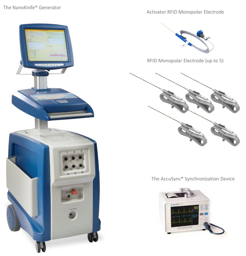
NanoKnife generator platform of the type used for irreversible electroporation procedures. Image: Guangzhou Fuda Cancer Hospital.
Two points matter for a technical reader. First, a single-needle configuration means one percutaneous access track rather than several electrode tracks; the report does not state the electrical parameters used, the pulse protocol, the delivery duration or the resulting ablation zone. Second, the report offers no comparative rationale against thermal ablation. A reader should therefore not infer that S-IRE was chosen over another modality for a documented technical reason. What is documented is the modality used, the anesthesia under which it was delivered, and the anatomy it was used in.
That distinction is the difference between a case report and a comparative study, and it is the reason this article treats NanoKnife cancer treatment here as a described procedure rather than as a demonstrated superiority claim.
Why biopsy was performed in the same session
The report lists concurrent biopsy among the procedures performed and pathology results among the deliverables. Combining sampling with the ablation session means tissue is obtained without a second puncture of a retroperitoneal lesion that is difficult to reach and sits against major vessels.
The practical value is downstream. In post-Whipple recurrence, pathology and molecular information is what later systemic decisions are built on, and having it available without a separate admission shortens the interval between the local procedure and the next planning step. The report does not state what the biopsy showed, whether it confirmed the recurrence histologically, or how the result altered subsequent management. Those are legitimate questions for a treating team and are not answered in the published case.
The role of the celiac plexus block
A celiac plexus block is an analgesia technique directed at the nerve bundle serving the upper abdomen, and the report records it as part of the same session alongside the ablation and the biopsy. Its function is pain control, not tumor control. In this case the report notes satisfactory pain control among the qualitative outcomes and describes the overall result as good recovery.
Placing the block in the same session has an operational logic that the report supports without quantifying: analgesia is established before the post-procedural period rather than after it, and the patient is already positioned for a percutaneous retroperitoneal approach. The report does not publish a separate pain score series, so the effect is recorded as a clinical impression rather than as a measured trajectory.
MDT evaluation as the decision gate
MDT evaluation precedes treatment selection in the workflow described by the hospital, and the pancreatic case follows that pattern. The sequence recorded is admission assessment, MDT review, procedural planning, the procedure itself, then imaging review and rehabilitation.
For a referring clinician or a patient reading this case, the MDT step is the part that is most transferable and least dependent on any single device. It is where imaging is read against the vessel anatomy, where anesthesia risk is weighed, and where alternatives such as interventional or systemic options are placed alongside local ablation. The report does not disclose the composition of the board or the alternatives discussed, but it does establish that the procedure was selected through a collective review rather than by a single operator.
What the outcomes show, and what they leave open
Two outcomes are stated in the report. The patient ambulated the next day, and CT showed evident necrosis and shrinkage of the lesion. Qualitative results are described as good recovery, satisfactory pain control and positive family feedback.
No numeric values accompany any of these statements. There is no pre- or post-treatment lesion measurement, no percentage of necrosis, no ablation volume, no length of stay in days, no follow-up duration and no survival endpoint. The CT images reproduced with the report are dated before treatment and the day after treatment, which is what allows the necrosis and shrinkage to be shown at all, but the images are not accompanied by measurements.
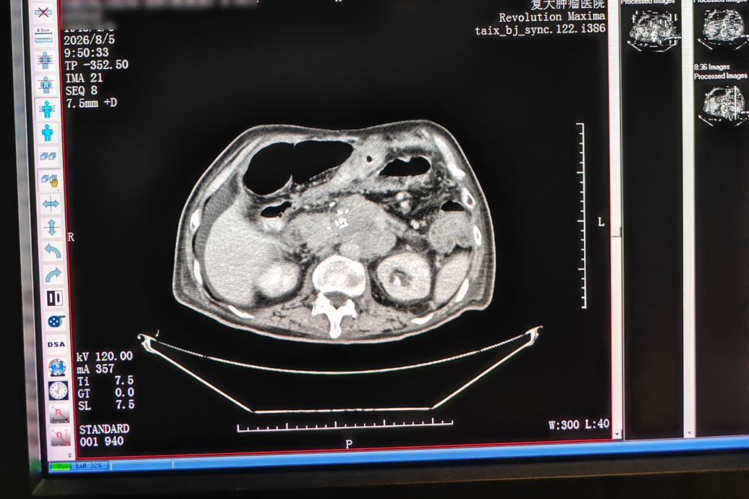
CT before treatment (5 August).
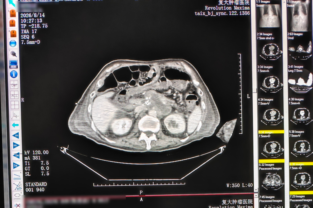
CT the day after treatment (14 August).
Imaging comparison reproduced from the case report. No numeric measurements were published with the images.
The honest reading is therefore narrow: in one 81-year-old patient with a vessel-encasing pancreatic recurrence, a single-needle IRE ablation under local anesthesia with dual-image guidance, concurrent biopsy and celiac plexus block was followed by next-day ambulation and CT evidence of necrosis and shrinkage, with no complications described in the report. Everything beyond that sentence requires evidence the report does not contain.
How the approach compares with conventional options
The comparison below is organized around what each option demands of the patient, because in this case the patient's tolerance was the binding constraint. It describes the documented case, not a trial result.
Table notes: the constraints in the third column are those recorded for the single reported patient, not general exclusion criteria. Thermal ablation is included as a category because it is part of the ablation family used in interventional cancer treatment; the case report does not compare outcomes between modalities.
The boundary that most needs stating is this: a single case report cannot establish that local-anesthesia S-IRE is preferable to any alternative. There is no control group, no comparison of anesthesia-related complications, no measure of local control over time, and no data on how often a lesion of this configuration can be treated this way. Its value lies elsewhere, in showing that a specific combination was executable in a patient for whom the conventional route was closed.
Where this configuration fits, and where it stops
The scenarios that the hospital describes for this family of approaches are unresectable tumors, locally advanced or recurrent disease, and organ-preserving treatment needs, including patients with limited benefit from traditional therapy and patients with inoperable tumors. The pancreatic case sits squarely inside that set: recurrent disease in a previously operated bed, vessel encasement, and a patient who could not undergo general anesthesia.
For a minimally invasive pancreatic cancer treatment evaluation, the case suggests a short list of questions worth putting to any treating team. Was the lesion assessed as non-resectable on imaging, and by whom? Was an anesthesia assessment documented before the modality was chosen? Was tissue obtained, and did pathology change the systemic plan? Was the procedure delivered in one session or staged? And what imaging follow-up schedule was set?
The place it stops is equally clear. The report does not define which pancreatic recurrences are suitable, does not describe a failed attempt, and does not address what happens when the lesion cannot be visualized adequately on ultrasound or when the patient cannot tolerate the procedure under local anesthesia. Minimally invasive treatment for metastatic cancer and for inoperable tumors is a growing field, but growth in the field is not evidence that any single configuration applies broadly.
Market context: expansion and the access gap
The wider market signal is one of steady expansion. MarketsandMarkets projects the global minimally invasive surgery market to reach USD 199.30 billion by 2030, from a 2025 base year. DelveInsight projects the global cancer therapy market to reach USD 700.09 billion by 2034. Grand View Research reports that Asia Pacific held a 37.6% revenue share of the cancer treatment facilities market in 2025.
Those figures describe capacity and spending, not availability at the point of care. The same body of clinical commentary notes gaps in the accessibility and indications of advanced techniques such as ablation, irreversible electroporation and CAR-T therapy, and restricted indications for unresectable tumors. Expansion at the market level and access at the patient level are different variables, and the difference is where cases like this one sit.
Two institutional signals are worth noting alongside the market data. Cryosurgery for cancer treatment was approved by China's State Drug Administration in 1999, which sets a regulatory baseline for ablation practice in the market where this case was treated. And in 2024, the hospital's research on cryoablation for lung nodules was cited in the expert consensus of the American Association for Thoracic Surgery, which indicates that institutional ablation work in this setting is being read beyond its home market.
What would move this from a case report to usable evidence
The next step is not a larger claim; it is a more standardized record. A registry-level dataset for image-guided ablation in pancreatic recurrence would need pre- and post-treatment lesion measurements, ablation zone volumes, anesthesia-related complications, pain scores, length of stay, time to ambulation, and follow-up duration with local control and survival endpoints. None of these are publicly available for this case, and hospital-specific outcome datasets of that kind are not generally published in this field.
If such reporting becomes routine, three things follow. Referring clinicians gain a basis for comparing local-anesthesia approaches against general-anesthesia ones rather than reasoning from anatomy alone. Procurement conversations shift from device specifications to outcome definitions. And case reports stop being read as proof of concept and start being read as entries in a series, which is what this one is best understood as today.
Frequently asked questions
Why was local anesthesia used instead of general anesthesia in this pancreatic S-IRE case?
The report states that advanced age and poor cardiopulmonary reserve made resection or general anesthesia intolerable for this 81-year-old patient, and that the percutaneous single-needle ablation was therefore performed under local anesthesia. The choice followed the anesthesia assessment rather than preceding it.
What does CT plus ultrasound guidance add during a single-needle ablation?
Ultrasound provides real-time visualization of the needle path during advancement, while CT provides the anatomical reference against the celiac trunk and superior mesenteric artery and verifies final position. The report describes this as real-time dual-image guidance and lists it among the services provided; it does not publish accuracy figures, needle-pass counts or radiation dose.
Why combine a biopsy with the ablation session?
The report lists concurrent biopsy among the procedures performed and pathology results among the deliverables. Performing it in the same session obtains tissue without a second puncture of a difficult retroperitoneal target, and pathology informs later systemic decisions. The report does not state what the biopsy showed or how it changed the treatment plan.
What is the purpose of the celiac plexus block?
The block is an analgesia procedure targeting the nerve bundle that serves the upper abdomen. In this case it was performed in the same session as the ablation and biopsy, and the report records satisfactory pain control as a qualitative outcome. It addresses pain rather than tumor control, and no separate pain score series is published.
What did the MDT evaluation decide before the procedure?
The workflow places MDT evaluation before treatment selection, and the case follows it: admission assessment, MDT review, procedural planning, the procedure, then imaging review and rehabilitation. The report does not disclose the board's composition or the alternatives it weighed, so the evaluation is documented as a decision gate rather than as a recorded rationale.
What outcomes does the case report document?
Two clinical outcomes are stated: the patient ambulated the next day, and CT showed evident necrosis and shrinkage of the lesion. Qualitative results are described as good recovery, satisfactory pain control and positive family feedback. No numeric values are provided for lesion size, ablation volume, length of stay, follow-up duration or survival.
Which details are case-specific and which are generalizable?
Case-specific: the 81-year-old patient's cardiopulmonary status, the approximately 7 cm lesion encasing both vessels, the post-Whipple anatomy, the specific anesthesia decision, and every outcome recorded. Potentially generalizable: the sequence itself, in which MDT evaluation precedes modality selection, dual-image guidance supports a percutaneous approach, and sampling and analgesia are integrated into one session. The report does not demonstrate that the sequence works the same way in other patients, and it should not be read that way.
Can this approach be compared with thermal ablation on the basis of this report?
No. The report documents the modality used and the anatomy it was used in, but gives no comparative rationale against thermal ablation and no comparative outcome data. Proximity to critical structures is a recognized selection challenge across ablation techniques, and this case illustrates one way of addressing it rather than a measured advantage over another way.
The hospital publishes an English-language brochure covering its treatment model, campuses and international patient services: Fuda Cancer Hospital brochure (PDF). General information is also available at www.fudahospital.com.
This article summarizes a single published case report and general industry context. It is not medical advice, and treatment decisions for pancreatic recurrence require assessment by a qualified multidisciplinary team.
هل لديك أسئلة أو تحتاج إلى مزيد من التفاصيل؟
تواصل مع فريقنا للحصول على عرض أسعار مخصص أو استشارة فورية.
طلب عرض أسعار
املأ النموذج أدناه وسيتواصل معك فريقنا بعرض مقترح مخصص.
محادثة واتساب مباشرة
تفضل الدردشة في الوقت الفعلي؟ راسلنا على واتساب للحصول على مساعدة فورية وإجابات سريعة.
- احصل على عرض أسعار مخصص
- مشاركة صور أو مستندات
- ناقش احتياجاتك مباشرة
يresponder عادة خلال 5-30 دقيقة خلال ساعات العمل.