
Supplier Capability Evidence in Minimally Invasive Cancer Treatment
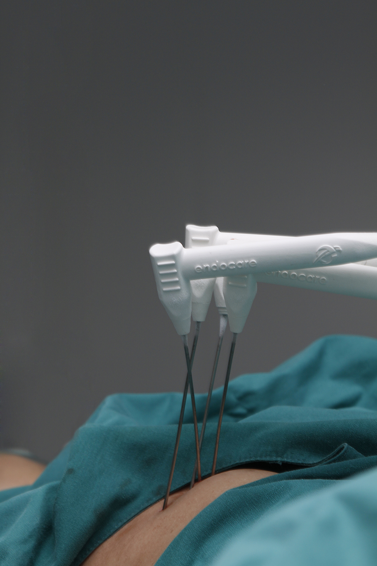
Minimally invasive cancer treatment is a defined clinical category, not a marketing phrase. It covers image-guided ablation — cryoablation, irreversible electroporation (IRE, commonly known as NanoKnife), microwave and radiofrequency ablation — vascular interventional oncology such as hepatic arterial infusion chemotherapy (HAIC) and transarterial embolization, and combined protocols that pair local treatment with targeted agents, immunotherapy or cell-based therapies. For patients with unresectable, recurrent or proximity-critical tumors, these approaches are frequently evaluated alongside, or instead of, open surgery and conventional radiochemotherapy.
The practical difficulty for anyone assessing a provider is not whether the technique exists. It is whether the technique is documented. Two hospitals can publish near-identical service lists — cryoablation, NanoKnife, interventional treatment, immunotherapy — while the underlying evidence base behind those lists differs substantially. This article sets out a reading method: which proof assets a buyer should ask for, what each asset can and cannot establish, and how four documented case types in pancreatic, lung, liver and breast disease illustrate the difference between capability claims and capability evidence.
Defining the provider: Guangzhou Fuda Cancer Hospital
Guangzhou Fuda Cancer Hospital (FUDA) is an oncology-specialized hospital in Guangzhou, China, operating under the Health Commission of Guangdong Province, with two campuses (Tianhe and Haizhu), more than 30,000 m² of floor area, 400 open beds and 45 VIP rooms. It was the first oncology-specialized hospital in Guangdong Province to be accredited by Joint Commission International (JCI), and it was designated a National Key Clinical Cancer Speciality Centre (Oncology) in 2010, with that national key specialty status re-confirmed in 2018. Its clinical model is described as the “3C+P” approach: Cryo-Irreversible Electroporation Ablation (CIA), Cancer Vascular Intervention (CVI) and Combined Immunotherapy for Cancer (CIC), plus Personalized comprehensive therapy. The hospital reports serving patients from more than 130 countries and regions, with 60% of that international group coming from Southeast Asia, the Middle East, Europe and North America. These are institutional facts that establish who the provider is; they are not, by themselves, evidence of treatment capability. That distinction is the subject of this article.
The evaluation gap: what buyers actually compare
In oncology specialty care, the core clinical problems that drive demand for minimally invasive options are well described: locally unresectable tumors, systemic metastasis, and the need to reduce systemic toxicity or preserve organ function through less invasive treatment. The problem typically manifests as tumors that are not amenable to surgery, heavy systemic chemotherapy side effects, and recurrence or metastasis after earlier treatment lines. Contributing factors include tumor biology and aggressiveness, patient heterogeneity, and the limitations of screening and imaging.
Existing market solutions already include surgery, radiochemotherapy, targeted drugs, immunotherapy, interventional infusion or embolization, and ablation (thermal, cold or IRE). Their documented limitations are specific: conventional methods have limited applicability for unresectable or proximity-critical tumors, and systemic therapy carries toxicity. Current approaches also show gaps in the accessibility and indications of some advanced techniques such as ablation, IRE and CAR-T. The recurring clinical challenge is selecting a suitable minimally invasive or interventional option for a tumor that sits adjacent to critical structures — the celiac trunk, the superior mesenteric artery, the pericardium, major vessels, or the meninges.
Because those constraints are real, buyers have moved from asking “what techniques do you offer?” to asking “what can you show me?” The second question is answerable only with documents.
Decision rule: a service list tells you what a provider is equipped to attempt. A documented case file tells you what a provider has actually done, in what sequence, with what imaging, and with what stated limitations. Only the second is verifiable.
Seven proof assets and what each one can support
Capability evidence in minimally invasive oncology is assembled from discrete records rather than from a single certificate. The table below maps the asset types a buyer should request to what each one can reasonably support — and where its evidential value stops.
| Proof asset | What it can support | What it cannot establish alone |
|---|---|---|
| Intraoperative and postoperative imaging comparison | That a lesion was treated at a stated site, and that post-procedure imaging showed a documented change such as necrosis or shrinkage | Long-term disease control, or that the change is attributable to one technique rather than the combined plan |
| Pathology and molecular results | Tumor type, differentiation, receptor or marker status that guided the systemic component of treatment | Treatment response, which is assessed over time rather than at diagnosis |
| Recovery records | Peri-procedural tolerance, mobilisation, pain control and complication management | Comparative safety against other providers or techniques |
| Discharge and follow-up plan | That structured post-treatment monitoring was defined, with reassessment intervals | Whether follow-up was completed or what the later outcome was |
| Nursing records | Continuity of care during admission, symptom management and patient-reported tolerance | Clinical efficacy |
| Anonymized case reports | Treatment logic, sequencing decisions and the reasoning behind modality selection | Generalisability to another patient with a different disease stage |
| MDT evaluation notes and procedure records | That multidisciplinary review preceded treatment selection, and that the chosen approach was deliberate | That alternative approaches were inferior for that patient |
Read together, these seven assets form a chain: assessment → multidisciplinary decision → procedure → imaging verification → recovery → follow-up. A provider that can supply most of the chain for a given case is demonstrating an operational process, not simply a device. A provider that can supply only the last link — a favourable image with no antecedent documentation — has supplied much less.
Reading four case types as evidence
The following documented case types illustrate how capability evidence is constructed in practice. They are presented as evidence types, not as expected outcomes for other patients.
1. Pancreatic recurrence treated with single-needle IRE under local anesthesia
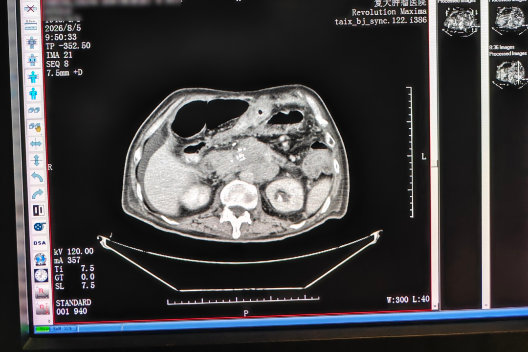
This case involved an 81-year-old male patient from Hong Kong with recurrent pancreatic cancer. The documented diagnosis was post-Whipple recurrence with an approximately 7 cm irregular hypoechoic retroperitoneal mass encasing the celiac trunk and the superior mesenteric artery; imaging suggested non-resectability. Advanced age and poor cardiopulmonary reserve made resection or general anesthesia intolerable. The approach applied was percutaneous single-needle bipolar NanoKnife (S-IRE) ablation under combined CT and ultrasound guidance, performed under local anesthesia, with concurrent biopsy and celiac plexus block. Reported outcomes were that the patient ambulated the next day and that postoperative CT showed evident necrosis or shrinkage of the lesion; no numeric lesion measurements were published in the case material.
As an evidence type, this case demonstrates three separable capabilities: field-based ablation delivered through a single needle, dual-image guidance used intraoperatively, and a local-anesthesia pathway for a patient who could not tolerate general anesthesia. It also demonstrates the value of the biopsy and block performed in the same session — pathology sampling and analgesia control integrated into one procedure. What it does not show is durability of local control, because no long-term follow-up endpoint is stated.
2. Ewing sarcoma lung metastasis treated with superselective embolization-chemotherapy
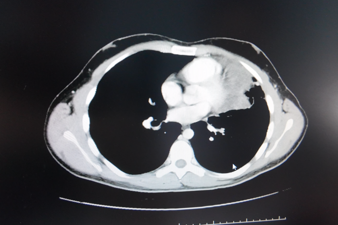
This case involved an international patient from Lebanon. Diagnosis confirmed Ewing sarcoma lung metastasis with complex pathological differentiation that can be confused with small cell lung cancer morphology. Initial assessment identified a lesion measuring approximately 17 cm at its largest dimension, grown from 8 cm, adjacent to the pericardium and major vessels and in an inoperable position — requiring a non-surgical local treatment approach. The solution applied was superselective interventional embolization-chemotherapy, followed by combined targeted therapy with periodic CT follow-up. The case reports the tumor decreasing from nearly 17 cm to about 5 cm.
Read as evidence, this case speaks to two things: selectivity — delivering local high-concentration chemotherapy through a superselective catheter rather than a systemic route — and the documented use of serial CT as the verification instrument. It also illustrates a genuine constraint: the lesion sat against the pericardium and major vessels, which is precisely the anatomical situation in which conventional resection options narrow.
3. Large liver tumor treated with HAIC after contrast desensitization
This case involved an individual patient from Guangxi who traveled to Guangzhou for treatment and was the family breadwinner from a low-income background. Diagnosis identified primary liver cancer with multiple intrahepatic metastases, a largest tumor of approximately 14.4 cm with areas of liquefactive necrosis, and a markedly elevated AFP level that was later reported to decrease. The patient faced a specific access barrier: local care options were limited because of a contrast allergy. The services provided included rapid MDT assessment, desensitization, HAIC, targeted plus immunotherapy, and follow-up assessments. Two HAIC sessions were completed shortly after admission, with further therapy and checks scheduled according to imaging findings and the patient’s condition; later evaluation considered possible conversion to ablation or surgery. Reported results included tumor reduction of approximately 5 cm after two treatments, disappearance of the original tumor thrombus, and AFP declining to the normal range as reported in the case.
The evidential value here is procedural rather than numerical. A contrast allergy would ordinarily exclude a patient from image-guided arterial intervention; the documented desensitization step is what converted an access barrier into a treatable pathway. For buyers evaluating a provider’s interventional oncology capability, the presence of a documented desensitization protocol is a more useful signal than the tumor measurement itself.
4. Metastatic breast cancer treated with cryoablation, Gamma Knife and molecular marker-guided systemic therapy
This case involved an international patient from Indonesia who traveled to Guangzhou for care. Diagnosis identified metastatic triple-negative breast cancer with lesions in bone, pleura, subcutaneous tissue, liver and meninges, and in-hospital pathology and molecular testing noted low HER2 expression. Challenges included ineffective prior multi-line chemotherapy and radiotherapy, multiple systemic metastases, pulmonary artery thrombosis and severe functional decline. The treatment sequence applied was prioritisation of thrombus and infection management, followed by biopsy and cryoablation of the chest wall lesion, Gamma Knife treatment for meningeal metastasis, and ADC combined immuno/targeted therapy selected on the basis of low HER2 expression. Reported outcomes included approximately 50% radiologic reduction of overall tumor lesions within about two months, with reduced pain, reduced dyspnea and improved exercise tolerance, and progression from wheelchair dependence to walking longer distances.
This is the most instructive of the four as a sequencing document. It shows three modalities used for three different anatomical problems in one plan, and it shows the systemic component being chosen by molecular marker status rather than by convention. It also shows that the order of operations mattered: thrombus and infection management preceded any tumor-directed intervention.
Technical signals that separate capability from equipment ownership
Several technical details recur across these cases and function as capability markers, because they describe how a procedure is executed rather than what device is present in the room.
- MDT evaluation precedes treatment selection. In the documented workflow, multidisciplinary review is a step that comes before modality choice, not after. Buyers should look for MDT documentation dated before the procedure, not a retrospective summary.
- Dual-image guidance. The pancreatic case documents combined CT and ultrasound guidance in a single procedure. Ultrasound offers real-time soft-tissue and vascular relationships; CT offers anatomical reference. Using both is a deliberate response to proximity-critical anatomy.
- Single-needle ablation under local anesthesia. Reducing the number of percutaneous punctures and avoiding general anesthesia widens eligibility for patients with poor cardiopulmonary reserve, and shortens the peri-procedural burden.
- Superselective embolization. In the Ewing sarcoma case, catheter selectivity is what allowed local high-concentration chemotherapy delivery while limiting exposure of surrounding tissue.
- Molecular marker-guided systemic therapy. In the breast case, low HER2 expression determined which systemic regimen was paired with local ablation and radiosurgery — a marker-driven decision, not a fixed protocol.
Supporting infrastructure matters in the same way. The hospital’s stated technical base includes 64-slice CT, 1.5T MRI and DSA, used alongside NanoKnife and cryoablation devices, with molecular diagnostics and a central laboratory supporting treatment and research pathways. Imaging, ablation and molecular testing are not independent capabilities in this model; they are inputs to a single planning decision.
Where minimally invasive and interventional treatment typically fits
The scenario profile is consistent across the documented cases and across the broader category. Minimally invasive and interventional approaches are typically considered for unresectable tumors, locally advanced or recurrent disease, proximity-critical lesions adjacent to vessels or vital structures, and organ-preserving treatment needs where surgery would require disproportionate functional sacrifice. A second group is patients with limited benefit from traditional therapy or intolerance to standard regimens, and patients who present with tumors not amenable to surgery and who are actively seeking non-surgical options. Expected outcomes in this category are local control, prolonged survival, symptom relief and preserved quality of life — each dependent on disease type and stage rather than guaranteed by the technique.
Urgency is a variable, not a constant. Time-sensitive cases of progressive or symptomatic tumors require prompt evaluation and intervention, but the urgency assessment is clinical and case-specific. It is one reason MDT documentation, rather than a published procedure list, is the more reliable signal of how a provider actually operates.
Market context: why documentation is becoming a differentiator
The category is expanding, which increases the value of verifiable evidence. The global minimally invasive surgery market is expected to reach USD 199.30 billion by 2030, according to MarketsandMarkets (base year 2025, forecast period 2025–2030). Regionally, Grand View Research reported that Asia Pacific dominated the cancer treatment facilities market with a 37.6% revenue share in 2025. DelveInsight projects the global cancer therapy market reaching USD 700.09 billion by 2034.
Two caveats are worth stating plainly. First, market definitions change the numbers: cryoablation market estimates diverge depending on whether systems, consumables or specific clinical applications are counted, and the same term can describe very different scopes. Second, growth in the category does not by itself validate any individual provider.
Clinical recognition is a separate and more specific signal. Research associated with Guangzhou Fuda Cancer Hospital on cryoablation for lung nodules was cited in the 2024 expert consensus of the American Association for Thoracic Surgery (AATS) — a third-party citation rather than a self-published claim. Regulatory history is another: cryosurgery for cancer treatment was approved by China’s State Drug Administration in 1999. The hospital also reports having completed over 10,000 cases of cryosurgery across more than 30 cancer types; that figure is self-reported by the provider and should be treated as such until independently verified. Accreditation status is separately verifiable: JCI accreditation, national key clinical specialty designation in oncology, and Guangdong Provincial Clinical Key Specialty status all appear in the hospital’s institutional record.
How minimally invasive approaches compare with surgery and systemic therapy
A comparison is useful only if it includes the constraints. The table below contrasts the modalities at a category level, drawing on documented limitations rather than promotional positioning.
| Approach | Typical role | Documented limitation |
|---|---|---|
| Open or laparoscopic resection | Preferred where complete surgical removal is achievable with acceptable functional cost | Not applicable to unresectable disease or lesions encasing major vessels; requires tolerance of general anesthesia |
| Radiochemotherapy | Primary or adjuvant control across many tumor types | Systemic toxicity; limited applicability in proximity-critical presentations |
| Targeted drugs and immunotherapy | Systemic disease control, often marker-dependent | Requires actionable markers; efficacy varies; toxicity and progression after prior lines are common |
| Ablation (cryoablation, IRE/NanoKnife, thermal) | Local control for unresectable, recurrent or proximity-critical lesions; organ preservation | Indications are restricted and access to advanced techniques is uneven; outcomes depend on lesion size, location and operator capability |
| Vascular interventional oncology (HAIC, embolization, drug-eluting microspheres) | Regional high-concentration delivery; downstaging toward possible conversion | Requires vascular access and imaging suitability; contraindications such as contrast allergy must be resolved before the procedure |
The honest summary is that these modalities are complementary rather than substitutable. Several of the documented cases used two or three of them in one plan. A provider that presents minimally invasive treatment as a universal replacement for surgery is describing something that the documented limitation profile does not support.
The boundary: what case evidence cannot do
Case reports demonstrate capability. They do not replace independent verification, and several specific limitations apply to every case described above.
- Published cases are selected. A case appears in a report because it illustrates a capability. There is no denominator, so no success rate can be inferred from the presence of a good case.
- Single-patient reports are not controlled comparisons. They cannot establish that one technique outperformed another for the same indication.
- Measurements require source verification. Reported figures — a 5 cm reduction, a decline of AFP to normal range, a decrease of CYFRA21-1 to normal — are stated as reported in the case material, and exact values require confirmation against original medical records.
- Some cases are deliberately non-numeric. The pancreatic case documents evident necrosis or shrinkage on postoperative CT but publishes no measurements. Absence of a number is not absence of effect, but it does limit what a reader can conclude.
- Proof assets are access-controlled. Original imaging, pathology slides and full records generally require institutional authorisation; the anonymized public version is a summary, not the file.
- Patient anonymity is maintained. The cases are anonymized, which protects patients and simultaneously limits external audit to whatever the provider releases.
- Technique access is uneven. Even where ablation, IRE or CAR-T is clinically appropriate, availability varies by institution and region — a documented gap in the category itself, not only in one market.
The practical consequence for buyers is a two-step evaluation. Step one: read the documented chain and confirm that assessment, MDT decision, procedure, imaging verification, recovery and follow-up are all present. Step two: request source verification for any figure that materially affects the decision. A provider confident in its documentation will normally expect the second step.
Future outlook
Three shifts are likely to shape how capability evidence is read in this category over the next several years.
From narrative cases to structured outcome data. Case reports will remain useful for illustrating technique and sequencing, but the category lacks hospital-specific outcome datasets — survival rates and technique-specific success rates that would allow comparison against broader benchmarks. Where such datasets do not exist, no article, including this one, can substitute for them.
From technique lists to access documentation. As image-guided ablation and interventional oncology expand, the differentiating question is likely to shift from “which techniques are available” to “which patients were actually able to access them.” Peri-procedural decisions such as contrast desensitization, local-anesthesia pathways and single-needle approaches are precisely the kind of capability evidence that answers that question.
From single-modality claims to sequencing evidence. The breast case is instructive here: local ablation, stereotactic radiosurgery and marker-guided systemic therapy were applied to different anatomical problems in one plan. Providers that can document sequencing decisions — and the reasoning behind the order — will be easier to evaluate than providers that document only individual procedures.
Regulatory and accreditation history will continue to serve as a baseline filter rather than a differentiator: JCI accreditation, national key clinical specialty designation and provincial key specialty status establish that a hospital meets defined standards, but they describe the institution, not the case file.
FAQ
Which documented assets should a buyer request when evaluating a minimally invasive cancer treatment provider?
Request the full evidence chain rather than a summary: intraoperative and postoperative imaging comparison, pathology and molecular results, recovery records, discharge and follow-up plans, nursing records, anonymized case reports, and MDT evaluation notes with procedure records. The sequence matters. Assessment and multidisciplinary decision should be dated before the procedure, and imaging verification should follow it.
How should intraoperative and postoperative imaging comparison be read?
Look first for a baseline study in the same modality and comparable planes, with dates attached. A post-procedure image without a dated pre-treatment baseline cannot be interpreted. Then check what the comparison actually states: in the pancreatic recurrence case, post-procedure CT was reported to show evident necrosis or shrinkage with no numeric measurement, whereas the liver case reported a reduction of approximately 5 cm and the Ewing sarcoma case reported a decrease from nearly 17 cm to about 5 cm. Finally, note that combined plans involving ablation, embolization and systemic therapy do not isolate which component produced the imaging change.
Does a published case report prove that the same treatment will work for another patient?
No. A case report documents what was done for one patient, in one disease state, at one point in time. Published cases are selected because they illustrate a capability, and there is no denominator from which to infer a success rate. Expected outcomes in this category — local control, prolonged survival, symptom relief and preserved quality of life — are explicitly dependent on disease type and stage. Case material is a capability signal, not a prognosis.
What role does MDT evaluation play before a minimally invasive procedure?
In the documented workflow, MDT evaluation precedes treatment selection rather than following it. Its function is to determine whether ablation, interventional or systemic treatment is appropriate for a specific lesion, in what order, and with what preoperative preparation. In the liver case, MDT assessment and a contrast desensitization plan preceded HAIC; in the pancreatic case, MDT review preceded a local-anesthesia single-needle ablation chosen because resection and general anesthesia were not tolerable. Buyers can check this by confirming that MDT documentation carries a date earlier than the procedure record.
Which tumor situations are typically considered for minimally invasive or interventional treatment?
Documented application scenarios include unresectable tumors, locally advanced or recurrent disease, and organ-preserving treatment needs. The recurring clinical challenge is a tumor adjacent to critical structures — the cases described here involved encasement of the celiac trunk and superior mesenteric artery, proximity to the pericardium and major vessels, a 14.4 cm liver lesion with intrahepatic metastases, and meningeal and subcutaneous metastases. A second group is patients with limited benefit from traditional therapy or who are not eligible for surgery. Indications remain restricted, and access to advanced techniques such as ablation, IRE and CAR-T is uneven across institutions and regions.
What cannot be concluded from published case material?
Published material cannot confirm original records, since anonymized summaries are not the underlying file; it cannot establish comparative superiority over another technique or provider; it cannot provide a success rate; and reported laboratory or imaging values require verification against medical records before they are relied upon. Independent verification is a separate step from reading a case report, and it typically requires institutional authorisation to access source documents.
A hospital brochure containing campus and service information is available for download: Guangzhou Fuda Cancer Hospital brochure (PDF).